

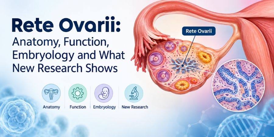
The rete ovarii is a small epithelial structure connected to the ovary that develops from mesonephric tissue. It is usually found around the ovarian hilum and medulla and has traditionally received far less attention than the ovarian follicles, stroma and other better-known reproductive structures. New research, however, is changing the way scientists think about its possible role.
In 2024, researchers published a more detailed account of the rete ovarii in the journal eLife and showed whether the structure is a secreting organ with links to the vascular, immune and nervous system of the ovary. They hypothesized it may play a role in ovarian homeostasis and development. Again, the work was carried out in mice so how this relates to human ovarian function is unknown.
What is the Rete Ovarii?
The rete ovarii is a network of epithelial cells and tubules associated with the ovary. It is considered homologous to the rete testis; the network associated with the male reproductive system.
The structure develops from cells of mesonephric origin, meaning it comes from tissue involved in the embryo’s early urogenital development. In adult ovaries, it is most commonly found near the ovarian hilum, although its extent varies between species.
The rete ovarii is easy to overlook because it is not the part of the ovary that produces eggs. It is a separate epithelial structure closely associated with the ovary.
Where is the Rete Ovarii Located?
The rete ovarii is primarily associated with the ovarian hilum, the area where blood vessels, nerves and other structures enter and leave the ovary. Some portions can extend into the ovarian medulla or into tissue outside the ovary.
Researchers describe three regions:
- Intraovarian rete (IOR): located within the ovary.
- Connecting rete (CR): links the intraovarian and extraovarian regions.
- Extraovarian rete (EOR): extends outside the ovary and forms a more developed tubular structure.
This three-part organization has become particularly important in recent research because the different regions appear to have different cellular characteristics.
What is the Function of the Rete Ovarii?
The exact function of the rete ovarii is still being worked out, but recent research suggests that it may have active roles in ovarian development, signaling and tissue maintenance.
For decades, the rete ovarii was often treated as a relatively unimportant developmental remnant. Earlier research had already suggested that it was not necessarily functionless, including observations of secretory material within its tubules.
The newer mouse study provides stronger evidence for biological activity. Researchers found secreted proteins inside the extraovarian rete and demonstrated movement of material through its lumen toward the ovary. The structure was also closely associated with blood vessels, macrophages and neuronal projections.
Scientists have therefore proposed that the rete ovarii could act as an auxiliary or signaling structure for the ovary. That is a developing area of research rather than an established clinical function in humans.
Is the Rete Ovarii a Remnant or a Functional Structure?
Current research suggests that calling the rete ovarii simply a functionless remnant may be too simplistic.
Its embryological origin has long been understood, but its role after development has been less clear. A 2025 review highlighted the growing evidence that the structure may participate in ovarian physiology and follicular dynamics.
The newer evidence changes the question from “Why is this structure still there?” to “What is it doing?”
Researchers have identified several clues:
- It remains structurally organized in adult tissue.
- Different sections have distinct cell types.
- Its extraovarian portion contains a lumen with secreted proteins.
- Material can move through that lumen toward the ovary.
- It is closely associated with blood vessels, immune cells and nerves.
- It may interact with processes involved in ovarian development and homeostasis.
What is the Rete Ovarii Made Of?
The rete ovarii is made primarily of epithelial cells arranged differently across its three regions.
The intraovarian portion can appear as solid cords of cells, while the connecting and extraovarian portions develop more recognizable tubular characteristics. The extraovarian rete contains epithelial cells, including ciliated cells, and ends in a dilated region in the mouse model studied by researchers.
This cellular variation is one reason researchers no longer treat the entire rete ovarii as a single uniform structure.
How Does the Rete Ovarii Develop?
The rete ovarii develops from mesonephric tissue during embryonic development.
The mesonephros is part of the embryo’s early urogenital system. Cells from this region migrate into and around the developing gonad and contribute to the rete ovarii.
Historically, this embryological origin has been one of the main reasons the structure has been compared with the rete testis. Both arise from related developmental tissue, although their anatomy and roles in female and male reproductive systems are different.
Research in mice has now provided a much more detailed picture of how the three regions of the rete ovarii form and remain connected.
Does the Rete Ovarii Have Anything to Do With Follicles?
Possibly, although the exact relationship between the rete ovarii and ovarian follicles is still being investigated.
The ovarian follicle is the structure that contains the developing egg and its surrounding support cells. The rete ovarii is not itself an ovarian follicle.
However, earlier research has connected the rete region with ovarian development, and recent reviews have raised the possibility that it could influence follicular dynamics.
The newer research is particularly interesting because it identifies the rete ovarii as a potential source of secreted factors. Whether those substances directly regulate follicle development in humans remains unanswered.
Can the Rete Ovarii Develop Cysts?
Yes, cysts arising from the rete ovarii have been reported, although they are uncommon.
Pathology literature describes rete ovarii cysts and cystadenomas, typically located in the ovarian hilus. A classic series reported 16 cysts or cystadenomas, two solid adenomas and one adenocarcinoma attributed to rete ovarii origin. These lesions were rare and required microscopic examination to distinguish them from other ovarian tumors.
This is different from saying that ordinary ovarian cysts are caused by the rete ovarii. Most ovarian cysts encountered clinically are not rete ovarii cysts.
The distinction is important because “rete ovarii cyst” is a specific pathological diagnosis, not another name for a common ovarian cyst.
Can Rete Ovarii Tumors Occur?
Rare tumors originating from or associated with the rete ovarii have been described, but they are uncommon.
Published pathology reports include benign adenomas and extremely rare malignant lesions. Because the rete ovarii sits close to other ovarian structures, identifying the precise origin of a lesion can require detailed histological and immunohistochemical examination.
Research has also examined a possible relationship between the rete ovarii and Brenner tumors. In one small series, the rete ovarii and the tumors had different immunohistochemical profiles, showing why their relationship cannot be established simply from their appearance under a microscope.
What Did New Research Discover About the Rete Ovarii?
The biggest recent development is evidence that the rete ovarii may actively communicate with its surrounding ovarian environment.
The 2024 eLife research used advanced imaging and proteomic analysis in mice to examine the structure in much greater detail than traditional microscopy allowed. Researchers identified three connected regions and found secreted proteins within the extraovarian rete.
The study also found that the rete ovarii is positioned alongside:
- Blood vessels
- Macrophages and other immune-related cells
- Neuronal projections
- Ovarian tissue
Together, these findings suggest that the rete ovarii could participate in communication between the ovary and its surrounding environment.
Does the Rete Ovarii Have a Known Function in Humans?
Not yet with the same level of certainty suggested by the recent mouse research.
The rete ovarii has been identified in humans and is recognized as part of ovarian anatomy. Earlier literature documented its structure and rare associated cysts and tumors.
However, the detailed functional experiments that revealed secretion, luminal transport and interactions with surrounding tissues were performed primarily in mice. The researchers themselves frame the findings in terms of potential ovarian function rather than a proven human mechanism.
That difference should remain clear when discussing the rete ovarii online.
Why is the Rete Ovarii Suddenly Getting More Attention?
Better imaging and molecular techniques have made it possible to study a structure that was previously easy to overlook.
The rete ovarii is not a newly discovered part of the ovary. It has been described for more than a century. What has changed is the ability to examine its cells, connections and secreted molecules in much greater detail.
The recent work has reopened questions about whether the rete ovarii contributes to ovarian development, follicular activity and tissue homeostasis. A 2025 review described this as a shift away from viewing the structure as biologically insignificant.
What we know versus what researchers are still investigating
|
Established or well documented |
Still being investigated |
|
It is associated with the ovary |
Its full physiological role |
|
It has mesonephric developmental origins |
Its contribution to human ovarian function |
|
It has intraovarian, connecting and extraovarian regions |
Whether its secreted factors directly affect follicles |
|
Rete ovarii cysts and rare tumors have been reported |
Its possible role in fertility and reproductive health |
|
It contains epithelial structures |
How its signaling system works in humans |
Why Does the Rete Ovarii Matter?
The rete ovarii matters because it may represent a previously underestimated part of ovarian biology.
For years, most attention has naturally gone to follicles, eggs, hormones and the ovarian stroma. The rete ovarii sat in the background.
New research does not mean that it has suddenly been proven to control fertility or cause ovarian disease. It means scientists now have stronger reasons to investigate what the structure actually does.
The most interesting unanswered question is no longer whether the rete ovarii exists. It is whether this small, overlooked structure helps the ovary communicate with its surrounding environment, and whether that function is conserved in humans.
This article is for educational purposes and does not diagnose ovarian conditions. A finding involving the rete ovarii on an imaging or pathology report should be interpreted by the appropriate gynecologist, radiologist or pathologist in its clinical context.